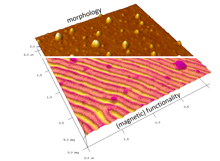
Since the introduction of the first commercial Atomic Force Microscope (AFM) in 1989, this technology evolved into an essential part in modern research and development. What started with imaging surface morphologies, expanded towards functional surface characterization with resolution capabilities down to the atomic level under special conditions. The possibilities to image surface properties in ambient, vacuum, and even liquid conditions are the key to use this unique technology from classical material sciences towards real time imaging of biological systems. In particular, AFM enables access to mechanical, electrostatic, magnetic, electric, thermal, optical or chemical information together with the 3D morphology (see right image). That perfectly complements other techniques such as Scanning Electron Microscopy (SEM), Raman Spectroscopy / Microscopy or Transmission Electron Microscopy (TEM) to provide comprehensive insights in material surfaces.
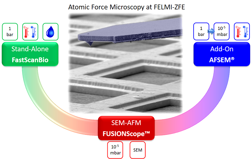
Following that motivation, FELMI-ZFE operates a variety of AFMs for different purposes. While all platforms can be used for classical material sciences, we also host a system, which allows operation in liquid environments with high speed capabilities at variable temperatures (FastScanBio). Furthermore, the first FUSIONScope™ was installed at FELMI-ZFE, which is a radically new, deeply integrated SEM-AFM platform for unique correlated microscopy studies. Finally, we have an add-on AFM (AFSEM®) for the flexible on-demand integration in other SEMs, Raman Microscopes and Focused Ion Beam (FIB) systems for a tailored workflow depending on individual requirements.