
Vibrational – & X-ray Spectroscopy

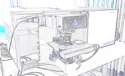
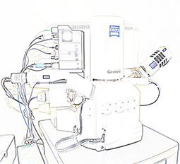
FT-IR Microscope Bruker Tensor 27 with Hyperion 3000

  • Bruker “Tensor 27” spectrometer with “Hyperion 3000” FT-IR microscope
  • Single element MCT detector
  • 64*64 elements FPA detector
  • 20x ATR (Ge crystal) objective
  • 20x Ref-objective with macro-ATR-stage (600×600 µm FoV)
  • GIR-objective
  • 16x transmission and reflection objective
  • “KnowItAll” software package for spectrum analysis
  • “OPUS” software package

Raman Microscope: LabRAM HR 800 with Olympus BX41

  • Confocal Raman microscope (Horiba Jobin Yvon)
  • Spectrograph with 800 mm focal length
  • Spectral resolution: 0,35 cm-1 (with 633 nm excitation and 1800 gr/mm grating)
  • Red laser: 633 nm, 17 mW
  • Blue laser: 473 nm, 20 mW
  • Green laser: 532 nm, 50 mW
  • BX41 microscope (10X, 50X, 100X objectives)
  • Motorised XYZ stage
  • 1024×256 pixel multichannel CCD detector (spectral range: 200–1050 nm)
  • LabSPEC software
  • Software module SWIFTTM for fast Raman imaging
  • DuoScan Imaging system (for fast and uniform scanning over small
    and large sample areas with variable pixel size)

Zeiss Sigma 300 VP

The Zeiss Sigma 300 VP represents the latest development in GEMINI® technology
and comprises a fully integrated inlens
detector (Inlens) for secondary electrons (SE) and an angle selective backscattered electron (HDAsB) detector.
The Sigma 300 VP offers ultra high resolution for both SE to image surface information and BSE to
present compositional information in the high vacuum mode. 

X-ray Diffraction

X-ray diffraction is widely used for crystallographic analysis to identify powders, thin films and crystals (e.g. minerals, inorganic compounds). Incident X-rays, in our case produced by a copper source, are diffracted into many specific directions when hitting the material. By measuring the angles and intensities of these diffracted beams, we are able to deduce the crystal structure, chemical bonding and various other information.