These substances are characterized by their high sensitivity to irradiation with high-energy electron beams. This is not an insoluble problem for us, however, because we have developed processes and equipment that enable investigation in micro- and nanoscopic size ranges. Many of the samples are also multicomponent mixtures and the boundaries between mineral fillers, layers of different compositions (layers) or mixtures of different phases of polymers. The explanation of some macroscopic phenomena by understanding the arrangements and peculiarities of these boundaries in the samples is indispensable for valid failure or damage analyses or for development of products.
Polymer & Soft Matter
How can we help you?
• Identification of Polymers (Granulates, Powders, Fibres)
• Defect Analysis, Quality Control
• Multi Layer Systems and Composites
• Morphology Studies (e.g. Phase Separation, …)
• Filler Particle Analysis and Agglomeration Studies
• In situ Experiments (Wetting, Tensile Testing)
• 3D Reconstructions
Your Contact Details

DI Dr. Armin Zankel
REM, EDX, 3D- und In situ-Tests
Tel. +43 316 873-8832
Contact me

Ing. Hartmuth Schröttner
Gruppenleiter REM/IR/Raman
Tel. +43 316 873-8349
Contact me
Morphology Study
With our microscopic methods, we also perform morphological investigations, such as phase separations, agglomeration behavior of additives and fillers. In addition to structural and morphological information, chemical information can also be obtained in high resolution due to the large pool of instruments.
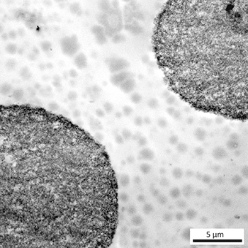
Additive Rich, Microphase Separated Polymer (Microtomy, TEM)
Material Identification
Combining SEM and Raman investigation in correlative experiments – we have such methods combined in one machine setup – allows to identify different materials – like filler particles (blue colored in the picture) and polymer phases (red colored in the image). Thus evaluation of filler-particle distribution and even particle size distribution becomes easily accessable.
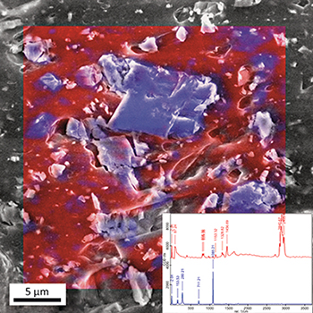
SEM-Raman: Correlative Microscopy of CaCO3 (Blue) in Polypropylene Matrix (Red)
Multi Layer System
By means of chemical imaging, it is possible to fall below the physical resolution limits (by diffraction of light Rayleigh criterion). This allows even very thin films to be examined with IR.
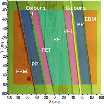
Cross Section Preparation FT-IR Spectroscopy: Chemical Imaging
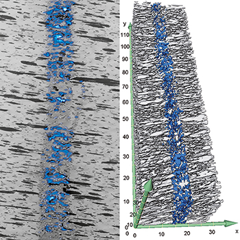
3D Reconstruction of Cracks
In situ wetting and tensile tests, ultramicrotomy-experiments (images of multiple layers prepared by microtomic cuts also in the electron microscope) and 3D reconstructions are also part of our portfolio. The determination of physical-mechanical parameters such as hardness, adhesion, deformation and energy dissipation is possible in micro- to nano-scale.
Cracks in Polymer after Tensile Test
In situ Ultramicrotomy in the ESEM