Paper is a very sophisticated product meeting different performance requirements. Therefore its composition is highly diverse and crucial to its properties. Its surface is conventially coated and contains various components. That is the reason why the detailled characterisation of paper remains challenging, especially on the microscopic level. We focus on new characterisation methods mostly based on electron microscopy but also on spectroscopic methods.
Paper and Fibres
How can we help you?
• Defect Analysis, Quality Control
• Multi Layer Systems and Interfaces
• Materials Characterisation
• Distribution of Filler Particles
• In situ Experiments (Wetting, Tensile Testing)
• 3D Reconstructions
• Structure Determination (Pulp, Fibres, Paper)
Your Contact Details

DI Dr. Armin Zankel
Key-Account-Manager – Papier
Tel. +43 316 873-8832
Contact me

Ing. Hartmuth Schröttner
Gruppenleiter REM/IR/Raman
Tel. +43 316 873-8349
Contact me

Ing. Claudia Mayrhofer
Präparation, Mikrotomie
Tel. +43 316 873-8347
Contact me
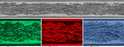
Elemental Composition and Mapping
by SEM-EDX
The detailed characterisation of the elemental composition helps to improve the understanding and reliability of various components to create valuable products. Analysis possibilities range from single, small particles to specific areas of interest and large area mappings (LAM) over areas in the mm range. The images show an LAM of an embedded paper and the results of EDX mapping indicating regions where the elements Calcium (Ca), Carbon (C) and Oxygen (O) were detected.
Correlative Morphology and Chemical Analysis
New instrumentation allows for the correlation of SEM and Raman microscopy. This method efficiently combines the high-resolution and morphological information (SEM) with chemical information (Raman) at the same sample position. The example below shows a cross section of coated paper (we use for posters at conferences) where the SE image is overlayed with a spectral mapping (red = epoxy resin, blue = cellulose, green = calcium carbonate, turquoise = filler particles).
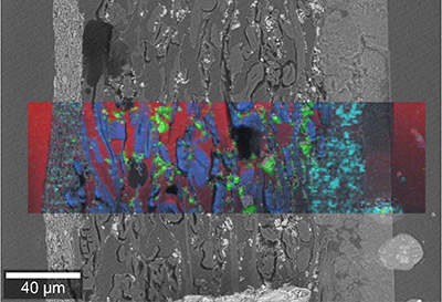
SEM-Raman analysis
Ca
C
O
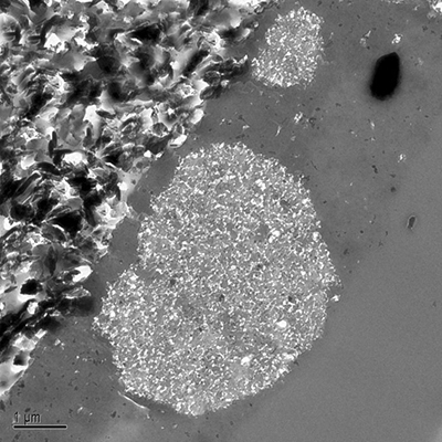
Multi-layer and Defect Analysis TEM
Detailed knowledge of defects like particulate impurities, black spots, fisheyes, gas bubbles helps to improve paper and ink quality. The TEM image below reveals that the printed ink agglomerates on the surface of the used paper.
Printability tester ”prüfbau”
Distribution of Filler Particles and Fibres: 3D Reconstructions
3D reconstructions are challenging. Nontheless they offer the most detailled insight into the distribution of different materials (filler particles, fibres, potential failures). The image below shows filler particles (grey) and fibres (green).
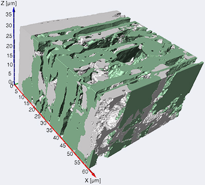
3D Reconstruction of paper